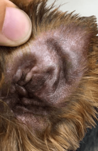
An aural (ear) hematoma is a blood-filled swelling on a pet’s ear. The condition occurs when blood vessels are broken, causing blood to pool between the skin and cartilage of the ear.
Causes of Aural Hematomas
Aural hematomas are typically caused by trauma to the ear. The condition is more common in dogs than cats, and is especially prevalent in pets prone to ear mites, allergies, and infections of the ear canal. Pets with these conditions often scratch their ears excessively and violently shake their heads. This leads to broken blood vessels in the ear and pooling of blood in the space between the skin and cartilage of the pinna (ear flap).
Symptoms of Aural Hematomas
Aural hematomas are characterized by a fluid-filled swelling on all or part of a pet’s ear. They can be firm or soft, and are usually warm and painful to the touch. Aural hematomas are often very irritating to pets, causing them to shake their head or paw at their ears continuously. Some aural hematomas may also be large enough to obstruct the ear canal.
Diagnosis of Aural Hematomas
Your veterinarian will ask you a series of questions to learn more about the onset of your pet’s symptoms. They will then perform a physical examination of the ear to check for underlying causes of the condition such as parasites, foreign bodies, or infection.
Other diagnostic tests will often be carried out to determine the cause of your pet’s aural hematoma. These may include routine blood work, urinalysis, and allergy testing. Advanced diagnostic tools such as X-rays and CT scans may also be used to check for signs of inner ear infection.
Treatment of Aural Hematomas
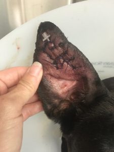
Treatment of aural hematomas involves draining fluid in the ear and promoting reconnection of skin and cartilage. The most effective way to achieve this is through surgical repair of the ear. While your pet is under general anesthesia, your veterinarian will make an incision on the inner surface of the ear along the hematoma. Fluid will then be drained and the ear flushed to remove any blood clots.
The next part of the surgery will involve reattaching the ear cartilage to the skin with sutures. The sutures will usually be left in place for several weeks while the ear heals. Your veterinarian may leave the incision open or insert a temporary drain into the ear. This will allow for continuous drainage as the wound heals. Other surgical methods may also be used to treat aural hematomas at the discretion of your veterinarian.
Following surgery, drugs may be prescribed to treat pain and inflammation. Other medications may also be necessary to treat the underlying cause of your pet’s aural hematoma.
Your veterinarian will advise you on keeping your pet’s ear clean and protected during the healing process. An Elizabethan collar (also known as an e-collar or cone) is an important part of treatment as it prevents pets from damaging their wound while it heals.
Prevention of Aural Hematomas
The best way to prevent your pet from developing an aural hematoma is to promptly treat conditions which can lead to self-trauma of the ears. These include ear infections, parasite infestation, and allergies. Scheduling regular checkups with your veterinarian is one of the simplest ways to minimize the risk of your pet developing conditions which can be a threat to their health.


