
Radiography is a common imaging technique that uses X-rays to examine tissues, internal organs, and bones. It is a useful tool that can help your veterinarian diagnose and treat many medical conditions in pets.
How Does Radiography Work?
Radiography machines project a beam of X-rays, a form of electromagnetic radiation, onto a photographic plate. When radiographs are performed, your pet is placed on the machine between the X-ray beam and the plate. X-rays penetrate structures within the body differently, depending on their density. They pass easily through the air and soft tissue but are mostly absorbed by denser materials like bone.
X-rays that pass through the body interact with the photographic plate below, producing an image. Because X-rays are mostly absorbed by dense materials such as bone, these areas will appear white on the X-ray image. Less dense materials including muscle and tissue will appear as shades of gray, while air cavities will appear black as they are least dense.
Is Radiography Safe?
Radiography is noninvasive and painless. It is also considered very low risk since low doses of radiation are used to produce X-ray images. If your pet is very anxious or in a lot of pain, it may be difficult for your veterinarian to position them correctly in the X-ray machine. In these cases, sedation may be required to obtain high-quality radiographs.
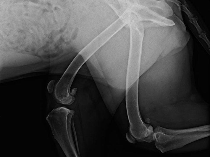
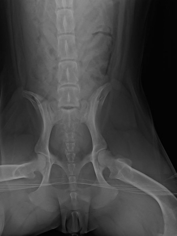
Hip, Knee, and Front Limb Radiography
Radiographs of the hips, knees, and front limbs can be used to assess trauma to the limbs and joints. They are also crucial for identifying conditions such as elbow dysplasia, hip dysplasia, or luxating patella.
If your pet has sustained trauma to the limbs, your veterinarian will check for evidence of fractures and bone damage. When orthopedic issues such as osteoarthritis or hip dysplasia are suspected, radiographs will help your veterinarian identify symptoms such as deterioration of cartilage and abnormal wear and tear on the joints.
Advanced imaging tools including magnetic resonance imaging (MRI) and computed tomography (CT) may also be used in combination with radiography to help your veterinarian visualize changes and damage to the joints.
Hip, Knee, and Front Limb Radiography Procedure
First, your veterinarian will measure the affected area of your pet’s body. They will then calibrate the radiography machine based on the measurements. Your pet will be placed on their side in the machine and X-rays will pass through them onto the photographic plate below.
Usually, your veterinarian will move your pet into a second position and repeat the procedure so that images can be taken from a different angle. Sedation is often required for radiographs of the limbs and joints since positioning your pet to obtain high-quality images can be painful or uncomfortable.
Image Gallery


